Home
/ How To View An Animal Cell Under A Microscope : Cheek Cells Under A Microscope Requirements Preparation Staining - Be careful pushing it under the clips that the cover slide doesn't move or crack.
How To View An Animal Cell Under A Microscope : Cheek Cells Under A Microscope Requirements Preparation Staining - Be careful pushing it under the clips that the cover slide doesn't move or crack.
How To View An Animal Cell Under A Microscope : Cheek Cells Under A Microscope Requirements Preparation Staining - Be careful pushing it under the clips that the cover slide doesn't move or crack.. The students learn how to make wet mount slides and focus them under the compound light microscope. Why does a specimen placed under the microscope have to be thin? Under a microscope, they look as though they're swimming through oil, neither very far, nor very fast. Finding mitochondria under a microscope download article. Cell membrane, cytoplasm, nucleus, dendrites, and axon.
The first reason is that plate cells are usually larger than their animal counterparts. Cheek cells under a microscope. There are millions of tiny cells to make up human being, but it will be painful to take out several cells in your hand or leg. Basophils do not ingest foreign cells. Show how to safely carry the microscope.
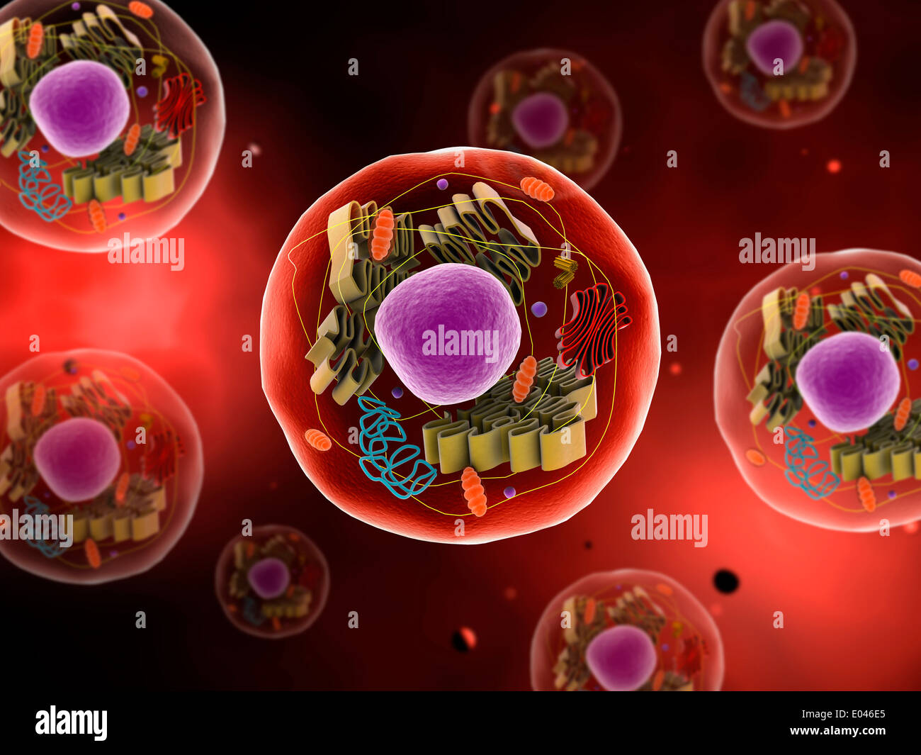
Outline Of Cell Biology Wikipedia from upload.wikimedia.org There's no set number of mitochondria in each cell, so it's hard to say how many you might see. Move the stage (the flat ledge the slide sits on) down to its lowest position. Actually the cells in your mouth can be taken out easily though it will be painful to take any cell out. Why does a specimen placed under the microscope have to be thin? Select the lowest power objective lens. Animal cell under a microscope. A generalised animal cell as observed under an electron microscope. See how a generalized structure of an animal cell and plant cell look with labeled diagrams.
Animal cells under a microscope.
A generalised animal cell as observed under an electron microscope. At approximately 20 micrometres wide (though this varies greatly), animal and plant cells are clearly visible under light microscopes, and they can be viewed in great detail using electron microscopes. Select the lowest power objective lens. Animal cell under a microscope. The first reason is that plate cells are usually larger than their animal counterparts. Megan thoemmes of north carolina state university in raleigh and her colleagues found, as had. Finding mitochondria under a microscope download article. Basophils do not ingest foreign cells. The two species live in slightly different places. Animal cells under a microscope. Turn the revolving nosepiece so that the lowest power objective is in position. To examine animals cells under a microscope. Grasp the arm with one hand and place the other hand under the base for support.
They contain granules filled with histamine, a substance this opens in a new window. Animal cells • there are a number of differences between plant and animal cells when they are viewed under a microscope • cell size and shape of animal and plant cells differ • some organelles are found only in one cell type, but not in both (cell wall and chloroplast in plant cells. A microscope is a gateway to a normally invisible world. Is your cheek cell an animal cell? However, it does not appear to be parapet wall bricks under the high power of microscope.
Gudu Ngiseng Blog Animal Cell Light Microscope from 2.bp.blogspot.com With light microscopy i can simply scrape some cells from my cheek smear them on a slide and look at them. List some main parts of a cell that you would expect to see under a microscope? Why does a specimen placed under the microscope have to be thin? Observation of animal cells 1. How do i stain/dye plant cells for viewing under a light microscope? Is your cheek cell an animal cell? Move the stage (the flat ledge the slide sits on) down to its lowest position. How many individual chromosomes are in one cell?
The most basic reason that cells are stained is to enhance visualization of the cell or certain cellular components under a microscope.
Interesting fact you're made from millions of tiny cells, but you can't take out a couple of cells in your hand or. There are millions of tiny cells to make up human being, but it will be painful to take out several cells in your hand or leg. Grasp the arm with one hand and place the other hand under the base for support. To examine animals cells under a microscope. Under a microscope, they look as though they're swimming through oil, neither very far, nor very fast. With light microscopy i can simply scrape some cells from my cheek smear them on a slide and look at them. Each of these epithelial cells was examined under the microscope as students. They contain granules filled with histamine, a substance this opens in a new window. Finding mitochondria under a microscope download article. 4 these same onion cells were viewed under a microscope which had not been adjusted how could the image have been adjusted and corrected, using what part of the microscope? Covers brightfield microscopy, fluorescence microscopy, and electron microscopy. In 2014, it became clear just how ubiquitous they are. The lesson will also take you through some exam questions on finding magnification using a scale and an image of a cell.
To view cells under a microscope, we need to make and prepare something called a specimen on a slide. In this lesson you will learn the method of how to view an animal cell under the microscope. Plant cells, animal cells and bacteria can be visualized through the light microscope. The most basic reason that cells are stained is to enhance visualization of the cell or certain cellular components under a microscope. How many individual chromosomes are in one cell?
Microscopic View Of Animal Cell Stock Photo Alamy from c8.alamy.com See how a generalized structure of an animal cell and plant cell look with labeled diagrams. (reproduced by permission of photo. Cheek cells under a microscope. Move the stage (the flat ledge the slide sits on) down to its lowest position. Each of these epithelial cells was examined under the microscope as students. How is it different from animal cell? They contain granules filled with histamine, a substance this opens in a new window. To examine animals cells under a microscope.
Draw and label the following representative parts of the neuron as seen under the microscope:
Epithelial cells surround the internal. If you meet some cell biologists and get them talking about what they enjoy most in their work, you may find it comes down to one thing: How many individual chromosomes are in one cell? The first reason is that plate cells are usually larger than their animal counterparts. 4 these same onion cells were viewed under a microscope which had not been adjusted how could the image have been adjusted and corrected, using what part of the microscope? Is your cheek cell an animal cell? Animal cell under a microscope. Cheek cells under a microscope. Grasp the arm with one hand and place the other hand under the base for support. Covers brightfield microscopy, fluorescence microscopy, and electron microscopy. View a prepared slide of nervous tissue under the microscope. Slides and how to estimate the size of a cell and how can we measure the size of a cell? When handling or carrying the microscope, always do so with both hands.
Share :
Post a Comment
for "How To View An Animal Cell Under A Microscope : Cheek Cells Under A Microscope Requirements Preparation Staining - Be careful pushing it under the clips that the cover slide doesn't move or crack."


Post a Comment for "How To View An Animal Cell Under A Microscope : Cheek Cells Under A Microscope Requirements Preparation Staining - Be careful pushing it under the clips that the cover slide doesn't move or crack."